Medical Science Meets Ocean Rescue: New Fellowship Bridges the Gap
How Biomedical Innovation Is Powering Marine Conservation
Crosscutting fellowship links ocean and human health
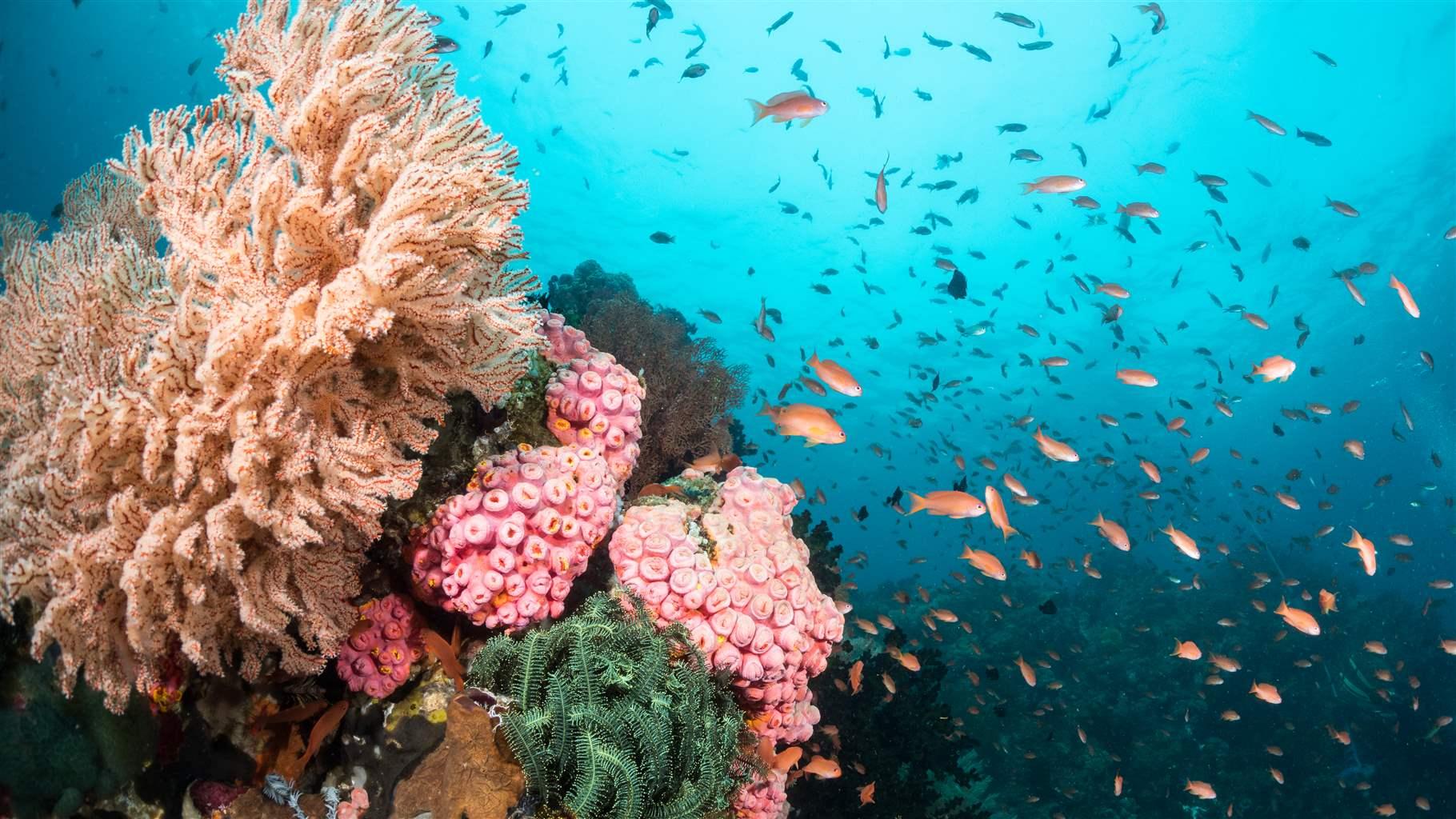
Human well-being and ocean health are closely intertwined: The marine environment plays a critical role in climate regulation and provides important resources to people—from food and medicines to nutritional supplements, coastline protection, and materials used in agriculture, cosmetics, and construction. But our oceans also face unprecedented threats, including pollution, overfishing, and climate change.
Marine conservation research is vital for finding solutions to these challenges and informing management decisions to protect ocean ecosystems. To strengthen connections between marine and biomedical sciences and foster innovative research with the potential to improve human health, The Pew Fellows Program in Marine Conservation and the Pew Scholars Program in the Biomedical Sciences have partnered with the Herbert W. Hoover Foundation on a fellowship positioned at the intersection of the two research fields.
Building bridges between disciplines
Much of the cutting-edge science being applied in marine conservation research today—including techniques such as gene editing and monitoring for environmental contaminants—has roots in the biomedical field, which is larger and typically better resourced. The new fellowship is designed to accelerate the transfer of techniques from biomedicine to marine science, with the goal of benefitting both people and the environment.
“We are thrilled to help launch this collaboration between the two Pew programs,” said Caiti Waks, program and outreach director for the Herbert W. Hoover Foundation. “Each program has a proven track record of identifying exceptional scientists and cultivating vibrant, collaborative networks within their respective communities. This crosscutting fellowship builds on those strengths by forging meaningful connections between the two programs.”
Supporting human and ocean health
Robert Richmond, a 2006 Pew marine fellow based at the University of Hawaii at Manoa, has seen firsthand the value of exchanges between marine science and biomedical research.
“Collaboration with colleagues in pharmacology has been critical to our success in advancing coral reef conservation,” Richmond explained. “By harnessing biomedical tools and approaches, we can now identify, understand, and address the sources of coral stress at sublethal levels—from chemicals found in common sunscreens to swimming pool water in runoff—and reduce them before corals lose reproductive capacity, bleach, or die.
“The crossover tools and techniques allow us to move from correlation to actual causation to diagnose and treat affected corals and evaluate the effectiveness of management interventions in real time, from days to weeks rather than months to years.”

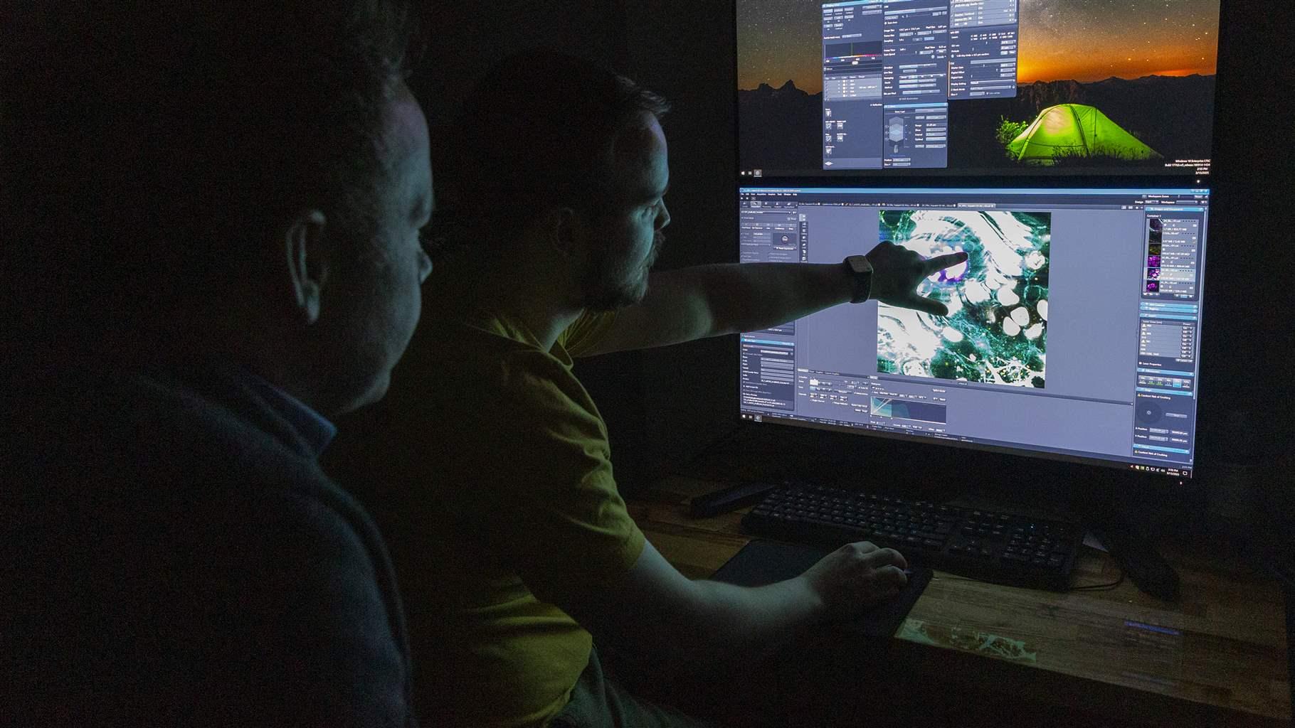
Tools and approaches such as advanced imaging and bioinformatics, which uses computers to analyze complex biological information, also have roots in biomedicine and are now commonly applied in marine science. These techniques have enabled researchers to evaluate the biodiversity effects of conservation interventions, such as oyster reef restoration, and to better understand and manage the population dynamics of vulnerable marine species.
Taking inspiration from the sea
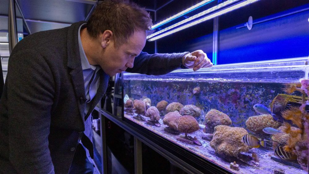
Phillip Cleves, selected in 2023 as the first recipient of the Pew-Hoover Fellowship in Marine and Biomedical Science, uses gene editing techniques to understand the factors that make some corals more likely to survive and recover from heat stress. He hopes to use this information to improve coral reef restoration by enabling practitioners to find wild corals with these traits so they can be propagated and protected—making reefs more resilient to climate impacts.
As a member of both Pew programs, Cleves has helped link the two robust, but often siloed, communities of scientific expertise.
“The [Pew-Hoover] fellowship presents the possibility of taking technology built for medicine and applying that technology to ecological problems, with the deep understanding that ecological health is human health,” Cleves said. “The connection between environmental and human health will only become more apparent as ecosystems degrade and pollution and populations increase, impacting our environment and contributing to human disease and suffering.”

Marine life such as corals and anemones can be useful model organisms for biomedical research, thanks to their extreme longevity and regeneration. But they remain a largely untapped resource that could help answer larger conservation questions as well as unlock new scientific insights that benefit both fields.
Accelerating progress in ocean protection
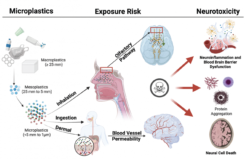
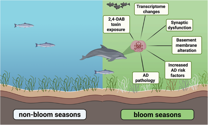
“The Herbert W. Hoover Foundation has a long history of supporting interdisciplinary work, particularly at the intersection of marine and human health,” Waks explained. “Recent HWHF-funded research identified links between harmful algal blooms in Florida and increased rates of neurodegenerative diseases in dolphins.”
“Subsequent studies in primates revealed similar patterns, demonstrating the neurotoxic effect harmful algal blooms can have on local populations,” she added. “Uncovering these connections is not only critical for protecting human health but also for inspiring public engagement. When people understand how environmental issues impact their own well-being, they’re far more likely to take action to protect both the environment and their health.”

“Ocean research has vast potential to benefit people—from drug discovery to improved environmental safety,” said Angela Bednarek, director of Pew’s scientific advancement portfolio. “The Pew Fellows Program in Marine and Biomedical Science is accelerating this type of interdisciplinary work to drive innovative solutions for human and ocean health.”
The next Pew-Hoover fellow will be announced in 2026.
Authored by Nathan Fedrizzi. Nathan Fedrizzi works on the Pew Fellows Program in Marine Conservation. To view the full article, please visit: https://www.pew.org/it/research-and-analysis/articles/2025/04/16/how-biomedical-innovation-is-powering-marine-conservation
How Nanoplastics Disrupt Cellular Membrane Structure
Nanoplastic-Induced Disruption of DPPC and Palmitic Acid Films: Implications for Membrane Integrity
With funding from the Herbert W. Hoover Foundation, Dr. Heather Allen of The Ohio State University published a study exploring how polystyrene nanoplastics—tiny plastic particles formed from the breakdown of larger plastics—interact with biological membrane components and potentially disrupt cellular function. The research demonstrated that nanoplastics can embed themselves into lipid films that mimic lung surfactants and cell membranes, altering their structure and potentially impairing critical biological processes like respiration, nutrient absorption, and cellular communication. These findings deepen our understanding of how nanoplastics may accumulate in living organisms and interfere with health at the molecular level. The study, conducted by Dr. Allen and her co-authors, is published in Environmental Science & Technology and can be accessed here (Allen et al., 2020).
Insights from Axolots into Human Spinal Chord Regeneration
Neuronal activation in the axolotl brain promotes tail regeneration
With support from the Herbert W. Hoover Foundation, Dr. Karen Echeverri of the Marine Biological Laboratory led a study published in npj Regenerative Medicine that uncovers a critical role for brain neurons in spinal cord regeneration in axolotls. The team’s findings reveal that neuron activation in brain regions distant from the injury site—not just in local tissue—plays a crucial, long-distance role in orchestrating regenerative repair through specific neuropeptides. This breakthrough challenges traditional assumptions that regeneration is driven only by cells near the wound and opens new avenues for exploring how the brain might influence healing processes throughout the body. The work holds potential implications for regenerative medicine, including the possibility of stimulating similar pathways in humans. The full study is available in npj Regenerative Medicine here.
Toxic Soil Discovered After Ohio Train Crash
Soil contamination by environmentally persistent free radicals and dioxins following train derailment in East Palestine, OH
With support from the Herbert W. Hoover Foundation, Dr. Joseph Ortiz and colleagues at Kent State University investigated the environmental and health impacts following the February 3, 2023, train derailment in East Palestine, Ohio. By analyzing soil samples collected near the derailment site months after the incident, the team found elevated levels of environmentally persistent free radicals (EPFRs) and dioxins/furans—harmful byproducts associated with combustion. Their findings provide the first field-based evidence that these toxic compounds may form together under real-world conditions, posing long-term risks to public and environmental health. This research underscores the need for ongoing monitoring, transparency, and remediation following industrial accidents, and contributes valuable insights into how complex chemical exposures may co-occur. The study is published in Environmental Science: Processes & Impacts and is available open access here (Lard et al., 2025).
Our Gut Mucus: A Shield Against Harmful Microplastics
The role of human intestinal mucus in the prevention of microplastic uptake and cell damage
With support from the Herbert W. Hoover Foundation, Dr. Ilana Brito at Cornell University led a study examining how human intestinal mucus serves as a barrier against ingested microplastic particles (MPs) and their damaging effects on gut cells. The researchers found that while the mucus layer effectively reduced particle movement, cellular uptake, and inflammation, certain types of plastic particles with specific surface coatings were able to penetrate the barrier and cause harm. These findings are critical in understanding how microplastics interact with the human body and highlight the protective role of mucus in gut health. The study also offers insights that could inform future research on exposure risks, microplastic toxicity, and even drug delivery technologies. The full study by Dr. Brito and her team is published in Biomaterials Science and is available open access here.
Spotting Harmful ‘Brown Tide’ Algae from Space and Shore
Field-Validated Detection of Aureoumbra lagunensis Brown Tide Blooms in the Indian River Lagoon, Florida, Using Sentinel-3A OLCI and Ground-Based Hyperspectral Spectroradiometers
With support from the Herbert W. Hoover Foundation, Dr. Joseph Ortiz of Kent State University and Dr. Edith Widder of ORCA led a study that successfully deployed a field-validated, hyperspectral imaging system to detect harmful Aureoumbra lagunensis (“brown tide”) blooms in marine environments. Calibrated across field sites and matched with lab-confirmed samples, their system demonstrated accurate real-time satellite identification of brown tide blooms in complex coastal waters. This work highlights the importance of combining advanced optical sensors with machine-learning classification to monitor harmful algal blooms in situ, providing environmental managers with a powerful tool for early detection and mitigation. The research by Ortiz, Widder, and colleagues is published in GeoHealth and can be accessed here (Ortiz et al., 2019)
Alzheimer’s Link? Toxic Algae Toxin Found in Brain Tissue
Detection of β-N-methylamino-l-alanine in postmortem olfactory bulbs of Alzheimer’s disease patients using UHPLC-MS/MS: An autopsy case-series study
With support from the Herbert W. Hoover Foundation, Dr. David Davis and a team of scientists at the University of Miami Miller School of Medicine contributed to a study exploring how exposure to airborne cyanobacterial toxins may be linked to neurodegenerative diseases like Alzheimer’s. The researchers examined postmortem olfactory tissue—located in the upper nasal passage—of individuals with varying stages of Alzheimer’s and detected the presence of BMAA, a neurotoxic compound produced by cyanobacterial blooms. The study raises important questions about whether inhaling aerosolized environmental toxins could play a role in triggering or accelerating neurological decline. By highlighting a potentially overlooked pathway of exposure through the nose, this work adds to growing evidence that environmental factors contribute to brain disease. The full study is published in Toxicology Reports and is available open access here.
Finding Nano Plastics in Seconds: A New Ultra-fast Test
NJIT Researchers Develop Rapid Method to Detect Micro- and Nanoplastics in Seconds
Microplastics and nanoplastics — tiny fragments shed from everyday plastic products — are increasingly found in our food, water, soil and even inside the human body. Their accumulation has been linked to fertility issues, metabolic disorders and other potential health risks in animal models. Yet detecting these pollutants has remained a time-consuming challenge.
To combat this challenge, researchers at New Jersey Institute of Technology and Rutgers have developed a powerful new method that can detect microplastics and nanoplastics in as little as 10 seconds, using a streamlined approach called Flame Ionization Mass Spectrometry (FI-MS).
The research, recently published in Journal of Hazardous Materials, shows that the technique can detect sub-microgram levels of plastic pollution in a wide range of samples, including bottled water, apple juice, agricultural soil and biological tissue. In one striking example, the team identified polystyrene nanoplastics in mouse placental tissue — a feat typically requiring complex chemical digestion and separation.
“It is amazing to have a method that can detect plastics in 1 milligram of tissue without time consuming sample prep,” said Genoa Warner, assistant professor in NJIT’s Department of Chemistry and Environmental Science and a co-author on the study.
Warner, who runs the Laboratory of Endocrine Disruption & Chemical Biology (EDC Lab), investigates the toxicity and mechanisms of endocrine disrupting chemicals in biological systems. She plans to use FI-MS to measure plastics in tissue samples.
“Traditional detection methods require hours or even days of sample preparation,” said Hao Chen, professor of chemistry and environmental science at NJIT, and lead author of the study. “With FI-MS, we can skip all of that. You can burn a dried sample — soil, tissue, even filter paper from water — and immediately detect trace plastics by their molecular fingerprint.”
The speed and simplicity of FI-MS could make it a valuable tool for both researchers and policymakers. While agencies like the EPA have yet to establish regulatory limits for microplastics in drinking water, rapid detection methods like this could help lay the groundwork for future public health standards.
“There are few techniques or methodologies currently available to accurately and consistently measure the micro/nanoplastic concentrations in environmental or biological samples,” said Phoebe Stapleton, co-author of the study and associate professor in the Pharmacology and Toxicology Department of the Ernest Mario School of Pharmacy at Rutgers University. “Understanding those concentrations is vital to studying the toxicology of micro- and nanoplastic exposures.
“Those tools that do exist are expensive to acquire and may be out of reach for many groups. This technique provides a viable alternative for the measurement of plastics.”
Unlike other mass spectrometry techniques, FI-MS uses a small open flame to simultaneously break down and ionize plastic particles at the point of analysis. The resulting ions are captured and analyzed by a high-resolution mass spectrometer, allowing researchers to identify the type and quantity of plastic without elaborate cleanup.
“Our method offers both speed and sensitivity, as it avoids sample loss and contamination during preparation,” said Mengyuan Xiao, a Ph.D. student in Chen’s research group. “And it also allowed quantitative analysis of microplastics and nanoplastics in soil and tissue samples.”
Chen’s lab has filed a patent for the FI-MS technique, and is currently exploring commercial applications with instrumentation partners. The group is also developing rapid methods for the detection of per- and polyfluoroalkyl substances (PFAS), another group of harmful environmental contaminants.
Looking ahead, the team plans to expand the method to analyze plastic accumulation in biological samples such as blood and brain tissue — work that could help illuminate the long-term health effects of microplastic exposure.
“The public doesn’t always realize how easy it is to ingest microplastics,” said Chen. “Using hot liquids in plastic bottles or heating food in plastic containers can release particles directly into what you eat. The more we can measure and understand these exposures, the better we can protect human health.”
This work is supported by the National Science Foundation, the National Institutes of Health, Herbert W. Hoover Foundation and an NJIT faculty seed grant.
This research was conducted by Chen’s group at NJIT (Mengyuan Xiao, Yongqing Yang, Terry Yu, Jerry Liu, Alex Guo), in collaboration with Warner’s group at NJIT (Hanin Alahmadi, Allison Harbolic, Genoa Warner) and Stapleton’s group at Rutgers University (Gina Moreno, Phoebe Stapleton).
The Hoover W. Hoover Foundation partially funded this project and innovation. For more information, please visit: https://news.njit.edu/njit-researchers-develop-rapid-method-detect-micro-and-nanoplastics-seconds
Microplastics Found in Newborn Organs: A Shocking Discovery
Startling Discovery: Scientists Find Microplastics in Infant Organs at Birth
The Herbert W. Hoover Foundation is proud to have funded this research project and look forward to all of the future advances in the field that result from it. Article originally appeared on SciTechDaily.org. Original article can be viewed here. The scientific article can be viewed here.
A Rutgers Health study has discovered aerosolized plastics in neonatal tissue following in utero exposure.
Plastic pollution, including tiny particles smaller than a grain of sand, has become ubiquitous, affecting even newborn rodents, according to a Rutgers Health study published in the journal Science of the Total Environment.
Researchers have long understood that micro- and nanoplastic particles (MNPs), which enter the environment through oxidation and natural degradation of consumer products, are easily deposited in the human body through inhalation, absorption, and diet.
Experts also understand that these pollutants can cross the placental barrier and deposit in fetal tissues.
What’s been unclear is whether these particles remained in tissue long after birth. The Rutgers Health researchers found that they do, at least in rats. Their data that could have implications for human health.
“Nobody wants plastic in their liver,” said Phoebe A. Stapleton, an associate professor of pharmacology and toxicology at the Rutgers Ernest Mario School of Pharmacy, and the study’s senior author. “Now that we know it’s there – as well as in other organs – the next step is to understand why and what that means.”
Research Findings in Rodents
To assess the persistence of micro- and nanoplastic particles in neonatal tissue following maternal exposure, Stapleton and colleagues exposed six rats to aerosolized food-grade plastic powder for 10 days during pregnancy.
Rodents are good test subjects for this type of study, Stapleton said, because humans and rodents both possess a hemochorial placenta, meaning that maternal and fetal blood don’t come into direct contact during circulation.
Two weeks after birth, two newborn rats – one male and one female – were tested for micro- and nanoplastic exposure. In both cases, the same type of plastic that the mothers inhaled during pregnancy were found in the offspring’s lung, liver, kidney, heart and brain tissue. No plastics were found in a control group.
Stapleton said the findings are one more piece of evidence illustrating the potential dangers of micro- and nanoplastics in the environment.
“These results raise concerns for the toxicological impacts associated with MNPs exposure, maternal-fetal health, and systemic MNPs particle deposition,” the researchers wrote.
Micro- and nanoplastics are a ubiquitous pollutant, and have been detected in food, farmland, seawater, and snow. Plastics have even been found in the world’s deepest oceans and highest mountains.
These invisible pollutants are potentially hazardous to human health. An increasing body of evidence suggests a strong correlation between micro- and nanoplastic particles and cancer, inflammation, impaired immune function, tissue degeneration, and cardiovascular problems.
Call for Further Research and Policy Action
Stapleton said she hopes her findings help create a sense of urgency among policymakers to make more funding available for research.
“Without answers, we can’t have policy change,” she said.
Eventually, the persistence of these materials in human tissue could lead to greater regulation, Stapleton said. While plastics have undoubtedly improved consumer products, too little is known about their long-term health impacts. As researchers fill knowledge gaps, regulators will be better equipped to protect public health, she said.
“I don’t think we’ll ever get rid of plastics altogether,” she said. “They’re too important for modern life. But I do think we might get to a point where we’ll have some policies to indicate which ones are less toxic than others.”
Reference: “Identification of micro- and nanoplastic particles in postnatal sprague-dawley rat offspring after maternal inhalation exposure throughout gestation” by Gina M. Moreno, Tanisha Brunson-Malone, Samantha Adams, Calla Nguyen, Talia N. Seymore, Chelsea M. Cary, Marianne Polunas, Michael J. Goedken and Phoebe A. Stapleton, 6 August 2024, Science of The Total Environment.
DOI: 10.1016/j.scitotenv.2024.175350
The research was funded in part by a grant from the Herbert W. Hoover Foundation.